Episode Details
Back to Episodes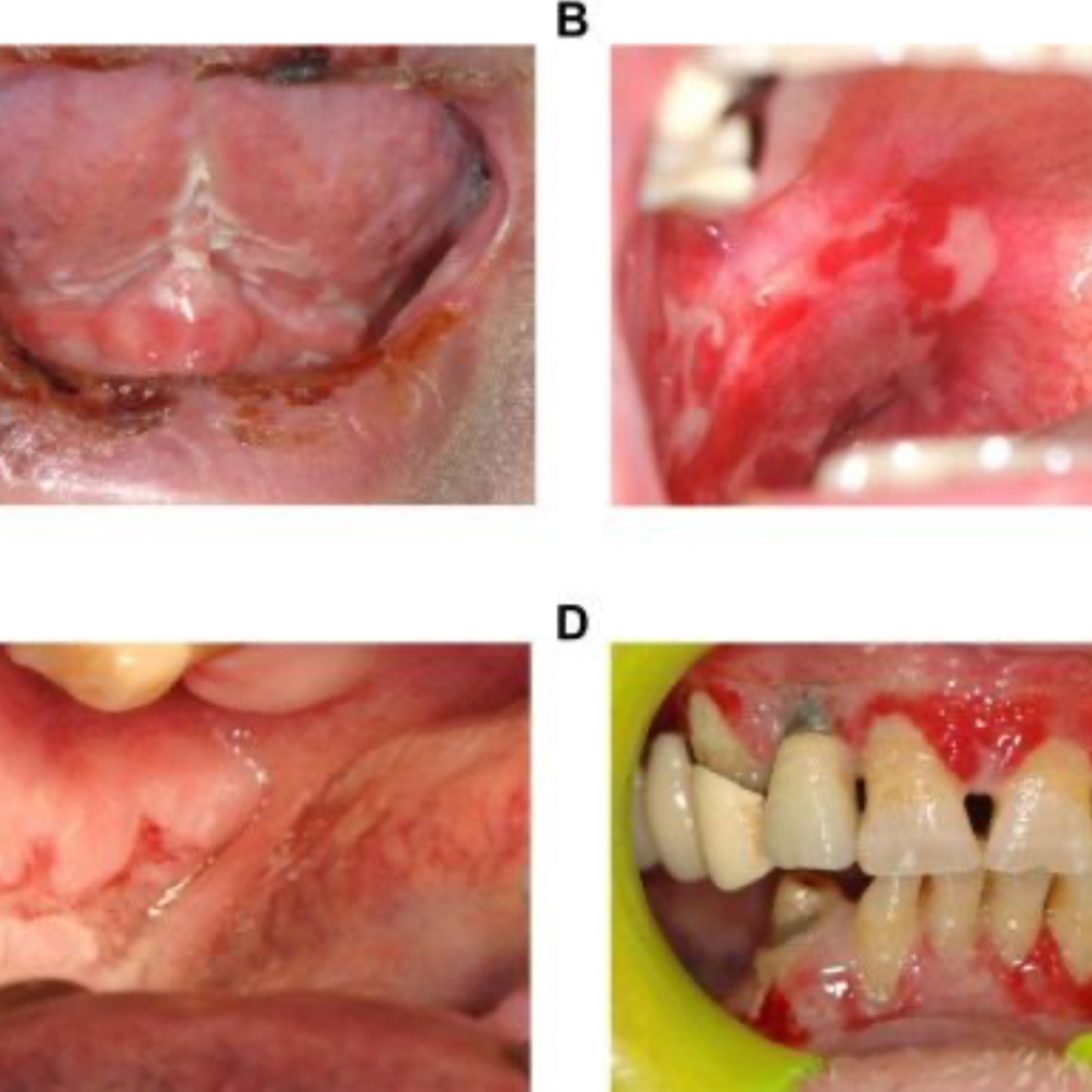
📝 “What are the Key Differences Between MMP, Pemphigus Vulgaris, EM, and Lichen Planus?”
Description
- 12.18.24
Quick Review #257 - #pathology #oralpathology #doctorgallagher #oralsurgery #oralsurgeon #dentist #dentistry #dental
1. Mucous Membrane Pemphigoid (MMP)
• Cause: Autoimmune; antibodies target hemidesmosomes (BP180, BP230) in the basement membrane.
• Clinical Features: Chronic blistering primarily involving mucous membranes (oral cavity, conjunctiva, nasal, and genital mucosa). Skin is less affected.
• Oral Presentation: Desquamative gingivitis and ulcerations. Lesions are often subepithelial, resulting in tense blisters that heal with scarring.
• Diagnosis: Direct immunofluorescence (DIF) shows linear IgG and C3 along the basement membrane zone.
• Prognosis: Slow progression but can cause significant scarring (e.g., blindness if conjunctiva is involved)
2. Pemphigus Vulgaris (PV)
• Cause: Autoimmune; antibodies against desmogleins 1 and 3, disrupting desmosomes.
• Clinical Features: Painful intraepithelial bullae that rupture easily, leaving flaccid erosions. Common on skin and mucous membranes.
• Oral Presentation: Early involvement of oral mucosa; erosions on buccal mucosa, palate, and gingiva. Positive Nikolsky’s sign (skin shearing with pressure).
• Diagnosis: DIF shows intercellular IgG deposition in a “chicken wire” pattern.
• Prognosis: Life-threatening if untreated due to widespread blistering and infection risk.
3. Erythema Multiforme (EM)
• Cause: Hypersensitivity reaction, often triggered by HSV infection or drugs (e.g., sulfonamides).
• Clinical Features: Acute, self-limiting condition with target lesions on skin. Severe forms like Stevens-Johnson syndrome involve mucous membranes.
• Oral Presentation: Painful ulcers and erythematous patches on lips, tongue, and buccal mucosa with bloody crusted lips.
• Diagnosis: Clinical; biopsy may show necrotic keratinocytes.
• Prognosis: Self-limiting, resolves in weeks. Severe forms require hospitalization.
4. Lichen Planus (LP)
• Cause: T-cell-mediated chronic inflammatory condition.
• Clinical Features: Purple, pruritic, polygonal papules with white striae (Wickham’s striae) on skin.
• Oral Presentation: Bilateral reticular form (asymptomatic white striae) or erosive form (painful ulcers). Common on buccal mucosa and gingiva.
• Diagnosis: Histology shows saw-tooth rete ridges and lymphocytic band infiltration at the basement membrane.
• Prognosis: Chronic but benign; erosive LP has malignant transformation potential
References:
1. Buonavoglia, A., Leone, P., Dammacco, R., Di Lernia, G., Petruzzi, M., Bonamonte, D., Vacca, A., Racanelli, V., & Dammacco, F. (2019). Pemphigus and mucous membrane pemphigoid: An update from diagnosis to therapy. Autoimmunity Reviews, 18(4), 349-358. https://lnkd.in/eYdYDkqW
2. Neville et al. (2022), Oral and Maxillofacial Pathology.
3. ChatGPT.2024.
#podcast #podcasts #dentalpodcast #dentalpodcasts #doctorgallagherpodcast #doctorgallagherspodcast #doctor #dentistry #oralsurgery #dental #viral #dentalschool #dentalstudent #omfs #surgeon #doctorlife #dentistlife #residency #oralsurgeon #dentist #doctorgallagher